Category: Food allergy
-
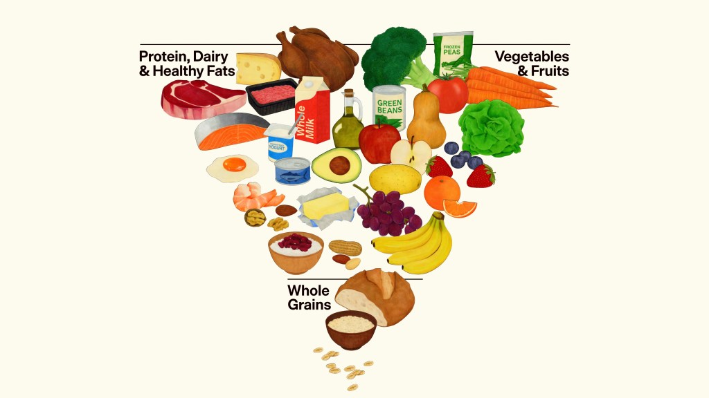
Expert Allergist Insights on RFK Jr.’s New Food Pyramid and Its Impact on Allergies
RFK Jr.’s new food pyramid has sparked debate regarding its implications for food allergies. The emphasis on protein in diets, especially for children, raises concerns as many allergenic foods are protein-based. While the pyramid encourages simpler, less processed foods, its effectiveness relies on consistent school communication and careful handling to manage allergy risks effectively.
-

Hidden Allergens in Popular Diets: What You’re Overlooking (and How to Fix It)
The shift toward diets like keto, vegan, and paleo introduces hidden allergens that can pose serious risks. Reformulations in packaged foods, cross-contact during food preparation, and mismanagement of allergies complicate safety. It’s essential to be vigilant about ingredient changes and maintain a food diary to distinguish between true reactions and dietary adjustments.
-

The Role of Vitamin D in Allergy Development and Prevention
Vitamin D deficiency is common and often overlooked in allergy patients, impacting immune functions and potentially worsening allergies. It regulates immune responses and supports gut health. Natural sunlight exposure is key for boosting levels, along with proper supplementation. Regular blood tests are essential for monitoring levels and ensuring optimal health.
-

6 Common Mistakes Parents Make With Food Allergens in Kids
Over 40% of children with food allergies experience severe reactions, often due to parental mistakes. Key errors include full-dose challenges, testing multiple allergens, neglecting investigation post-reaction, over-reliance on blood tests, and inadequate food safety.
-

How AI Is Revolutionizing Food Allergy Diagnostics & Healthcare
Imagine a world where diagnosing food allergies is precise, fast, and accessible to everyone. That future isn’t far off thanks to artificial intelligence (AI). With its unmatched ability to process and analyze vast amounts of data in seconds, AI is set to transform how we diagnose and manage food allergies. As a practicing allergist with…
-
6 Food Allergy Myths That Could Put You at Risk
Food allergies are often misunderstood, and misinformation can lead to dangerous assumptions. Whether you’re managing allergies yourself or looking out for a loved one, it’s time to separate fact from fiction. As an allergist doctor with over thirty years of experience, here are six of the most common myths about food allergies I see—and the…
-

Understanding Skin Testing Methods for Allergies
Which one (skin testing or blood testing) should I do?
-

Do What We Do Best! Desensitize
Tired of treating only your allergy symptoms and not getting to the root cause?
-

Food Allergy Reality Check – Distinguishing Between Fact and Fiction-Unraveling the Truth Behind the Hype
Thanks to YouTube, I present an excellent 9:20 on food allergy myths from 2 New York allergists. See the end of this post for their presentation–I hope you enjoy it as much as I did. In recent years, the conversation surrounding food allergies has significantly intensified. With constant news headlines, social media discussions, and shared…
-

Food allergy is making a run for it
For those hoping for an answer to their food allergies, experts have recently shared a glimmer of hope. Food allergy research has gained momentum and is advancing at astonishing rates compared to the advancement in studies on hayfever. Recent years have seen increased collaboration between universities and other medical organizations working together towards unraveling the…
